
Insights
From stratospheric antennas to implantable bio-sensors, from the thermodynamics of carbon capture to quantum technology and AI-designed biology, we use deep tech to challenge and change the status quo.
Read our latest research and insights to help you see beyond the innovation hype and find out how emerging technologies are solving important business problems.

Human-centred design innovation wins gold at iF Design Awards
Cambridge Consultants wins three iF Design Awards, including Gold, for human-centric design alongside Apple and Google ...

Rethinking diagnostics in the fight against antimicrobial resistance
Alejandra Sanchez and Ben Wicks explore why primary care needs timely, pragmatic insight, not lab-grade certainty, to enable better antibiotic decisions and stewardship ...
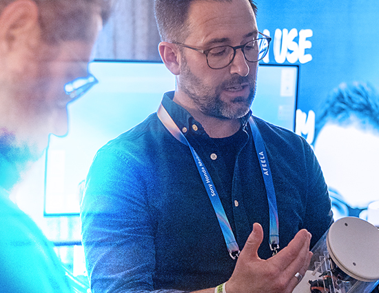
Smaller, smarter, even more radical – my post-CES vision for consumer innovation
First-principle science and engineering can lead to smaller, smarter and even more radical first-of-its-kind consumer products and commercial advantage ...

AI-powered networks are coming – telecoms must choose its path now
In CC’s paper developed with Verizon, we explore the shift to AI-powered networks and what telecom operators must do now to lead, not follow ...
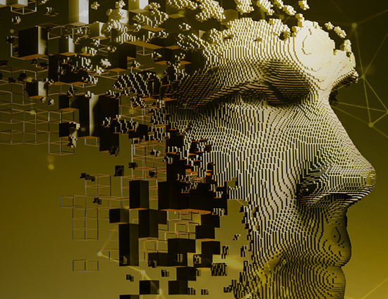
How we’re taking physical AI from simulation to reality
As we bring embodied AI into the real world, we’re accelerating the path to deployment while ensuring trust, reliability and impact through Sim2Real ...

Mastering fine manipulation for physical AI
Fine manipulation will be key to unlocking physical AI’s value, enabling humanoid and intelligent robotics to understand and act reliably in the real world ...

Telecoms can become the backbone of physical AI, or miss their moment
Discover the opportunity physical AI offers for telecoms leaders to harness edge AI and private 5G for real, scalable robotics ...
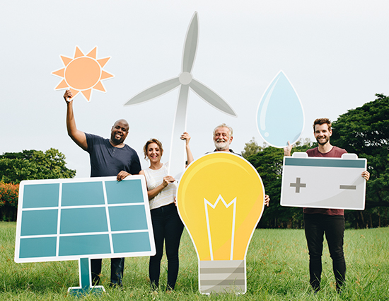
Our collaboration with Northern Powergrid helps drive the urban Community DSO smart energy initiative
CC’s Cloud Energy platform is being deployed by Northern Powergrid for their Community DSO project. Project leaders expect the trials to demonstrate how by working together, communities can help lower their peak energy consumption, benefiting the electricity network and the ...

Building trust into physical AI
Our AI assurance policy helps industry leaders understand, contextualise and manage risk to enable responsible physical AI deployment and innovation ...